MRI atlas of the human cerebellum / [edited by] Jeremy D. Schmahmann [and others].
Material type: TextPublication details: San Diego : Academic Press, 2000.Description: 1 online resource (xiv, 167 pages) : illustrations (chiefly color)Content type:
 - text
- computer
- online resource
- 9780080574073
- 0080574076
- 1282167480
- 9781282167483
- 9786612167485
- 6612167483
- 611/.81 22
- QM455 .M75 2000eb
- 2000 I-522
- WL 17
- digitized 2010 HathiTrust Digital Library committed to preserve
| Item type | Current library | Collection | Call number | Status | Date due | Barcode | Item holds | |
|---|---|---|---|---|---|---|---|---|
 eBook
eBook
|
e-Library | EBSCO Medical | Available |
Includes bibliographical references (pages 19-20).
Print version record.
Front cover; Title page; Copyright page; Dedication; Table of contents; Foreword; Preface; Acknowledgements; Chapter 1: Introductory text; Chapter 2: Images.
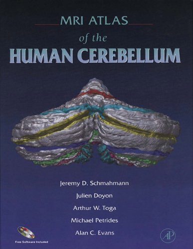
The MRI Atlas of the Human Cerebellum constitutes the most complete, detailed work on the human cerebellum to date. This definitive work provides images in the three cardinal planes (sagittal, transverse, and coronal) at closely spaced intervals of 2 millimeters. The images are derived from MRI scans of one individual and from postmortem sections of another. It is the only such atlas set within the universally accepted framework of the Talairach stereotaxic system, derived from standard landmarks in the brain. The book includes a new nomenclature system (labeling system) which is easier to use.
Use copy Restrictions unspecified star MiAaHDL
Electronic reproduction. [S.l.] : HathiTrust Digital Library, 2010. MiAaHDL
Master and use copy. Digital master created according to Benchmark for Faithful Digital Reproductions of Monographs and Serials, Version 1. Digital Library Federation, December 2002. MiAaHDL
http://purl.oclc.org/DLF/benchrepro0212
digitized 2010 HathiTrust Digital Library committed to preserve pda MiAaHDL
English.
WorldCat record variable field(s) change: 650